Colour Phase Contrast with a simple condenser | |
As a consequence of the relatively large (approx. 30mm) diameter of its transmitted light illuminating beam, the Zeiss Ultraphot can provide a simple and cheap means of producing colour phase contrast – without the need for a phase contrast condenser. Apart from standard ‘Phase 2’ objectives, the only major component required is one of the ‘flip-top’ Abbe-type condensers, modified by the removal of its upper lens and iris diaphragm assemblies. What then remains is a single lens condenser of about 24mm clear aperture and 35mm focal length. A Rheinberg-type disc of 32mm diameter is then prepared, with a central patch stop of 24mm. (I mostly use polyester lighting gel material from Strand Lighting or Lee Filters sample swatches, making both cuts at the same time (fig 1), using a concentric wad punch tool (fig 2), and a lump hammer on the end-grain face of a block of beech,). As is usual with Rheinberg discs, the patch stop and annulus should be in strongly contrasting colours, and the light transmission of the latter, three or more times that of the former. The completed disc is then placed on, say, a 32mm ‘daylight’ filter as a support, and dropped into the filter-well in the base of the microscope. This disc acts as an effective illuminating annulus for Ph2 objectives, other than (for some unknown reason) the 16/0.35, with which it gives an unevenly illuminated field of view. With the objective focused on, say, a diatom preparation, and the Bertrand lens swung in, the condenser is then raised or lowered until the image of the patch stop just fills the central area of the rear aperture of the objective as far as the inner margin of the ring of the phase plate, which itself is illuminated by the annulus of the Rheinberg disc. (An absence of gaps between the patch stop and annulus of the Rheinberg disc is absolutely essential, and concentric punches appear to provide the only ‘low-tech’ means of achieving this.) | |
 Fig. 1. |
 Fig. 2. |
A less demanding technique, allowing some inaccuracy in the cutting of the 4mm-wide annulus (not to mention ease of manipulation) requires only the annulus itself to be in the filter well, and the patch stop, in this case only 16mm in diameter, is placed on the upper surface of the condenser. Then, with the Bertrand lens in position, the stop is simply centred with the fingers, and the condenser focused so that the image of the stop fills the central area of the BFP of the objective, as before. A slight amount of overlap by the image of the annulus appears to be of little importance. In a more sophisticated version, a 24mm disc of ‘Polaroid’ material is used as a patch stop within the annulus in the filter well. Then, as long as the 16mm stop on the condenser is not anisotropic (e.g. it is cut from ‘gelatine’ rather than polyester sheet), a rotating analyser can be used to vary the brightness and saturation of the background of the image. This method will also (very usefully) reveal birefringent components in the specimen. | |
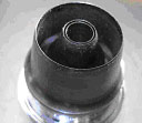 Fig. 3. |
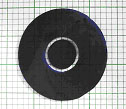 Fig. 4. |
Apart from their possibly enhanced appeal to publishers’ Picture Researchers, the images yielded by monochromatic colour phase contrast appear to show little advantage over those produced with orthodox equipment. However, the effect of using the two-colour system on diatoms, spicules, etc, in resin; and Protozoa, rotifers, etc. in water, can be quite spectacular, being initially more-or-less indistinguishable from that of Rheinberg illumination, but with the signal advantage of being easily produced with objectives of higher aperture. More importantly, careful examination of the image will show that there is a fairly typical phase contrast image superimposed on it, revealing considerable additional detail. More weakly retarding specimens produce a conventional phase contrast effect in shades of the 'background' colour, while the other hue is confined to areas that would normally show phase reversal, including out-of-focus objects. Thus when a disc with a dense blue stop and yellow annulus is used with a wet ‘cheek scrape’ preparation, the squames, leucocytes, and bacteria appear in mostly in shades of blue, but their haloes appear (pale) yellow. BIBLIOGRAPHY Bennett, A.H., Osterberg, H., Jupnik, H., & Richards, O.W., 1951, Phase Microscopy. Wiley & Sons. Grigg, F.C., 1950, Colour-Contrast Phase Microscopy, Nature, Vol., 165, no.4192 van Wesel, R., March 2004, Colour Phase Contrast. Micscape Magazine (www.microscopy-uk.org.uk) | |